
Our Radiology Doctors
Specialist doctors serving in our Radiology department

Diagnostic imaging services, including the performance and reporting of investigations, are provided.
In our Radiology department, diagnostic investigations are performed and reported using various medical imaging methods. The department works in coordination with other clinical units to support the diagnostic process. Each investigation is planned according to the patient's clinical status and the physician's request.
General assessments, routine screenings and diagnostic investigations are offered using digital X-ray, ultrasound and other imaging methods for different purposes. During the reporting process, clinical findings and imaging results are evaluated together. The patient is informed about the process both before and after the procedure.
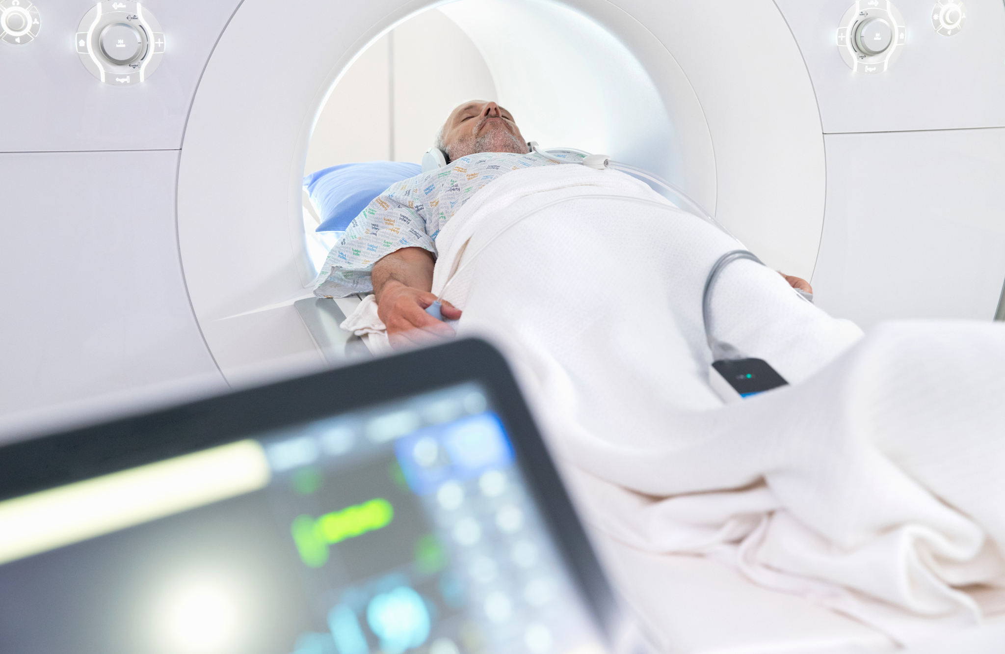
Magnetic Resonance Imaging (MRI)
Magnetic Resonance Imaging (MRI) is a non-ionizing, radiation-free diagnostic technique that produces high-resolution images of the body's soft tissues, organs, vessels and joints using a strong magnetic field and radio waves. Our department offers both Open MRI and Closed (1.5 Tesla) MRI options.
Areas and Conditions Imaged with the 1.5 Tesla MRI
Our 1.5 Tesla scanner operates at the magnetic field strength considered the gold standard for clinical diagnosis. The following regions can be examined in detail:
Brain and Nervous System
Stroke diagnosis and follow-up
Brain tumors and masses
Multiple sclerosis (MS) plaques
Dementia and Alzheimer assessment
Aneurysms and vascular pathologies (MR angiography)
Pituitary gland evaluation
Epilepsy investigation
Investigation of headache and dizziness causes
Spine and Spinal Cord
Lumbar and cervical disc herniation
Spinal stenosis
Spinal tumors
Post-traumatic spinal cord injury
Scoliosis assessment
Ankylosing spondylitis
Joints and Musculoskeletal System
Knee: Meniscal tears, anterior/posterior cruciate ligament injuries, cartilage damage
Shoulder: Rotator cuff tears, labrum injuries, frozen shoulder
Hip: Avascular necrosis, labral tears, femoroacetabular impingement
Ankle and wrist injuries
Bone tumors and infections
Muscle tears and soft tissue lesions
Abdomen and Pelvis
Liver masses and hepatic steatosis
Biliary system imaging (MRCP — magnetic resonance cholangiopancreatography)
Pancreatic diseases
Kidney masses and adrenal gland evaluation
Uterine, ovarian and gynecologic pathologies
Prostate evaluation (multiparametric prostate MRI)
Bladder conditions
Vascular System (MR Angiography)
Neck vessels (carotid arteries)
Brain vessels
Abdominal aorta and renal vessels
Peripheral vascular disease (can be performed without contrast)
Breast
Breast MRI screening for high-risk patients
Evaluation and characterization of suspicious masses
Open MRI
Open MRI is a magnetic resonance imaging method performed with a wide, open-bore system. Unlike the narrow tunnel of conventional units, it has a wide and open structure. This makes it a more comfortable option for the following patient groups:
Patients with claustrophobia
Larger / obese patients who cannot fit into closed MRI units
Pediatric patients (a parent can stay nearby)
Elderly patients and those who have difficulty remaining still
Patients with disabilities or special needs
Powered by a strong magnetic field, it provides diagnostic-quality images and is used across a wide range of areas including the head, spine, joints (knee, shoulder, hip), abdomen and pelvis.
Closed MRI (1.5 Tesla)
Closed (conventional) MRI is the standard magnetic resonance examination performed with a high-field cylindrical scanner. Our scanner's 1.5 Tesla magnetic field strength provides higher image resolution, shorter scan times, and the ability to obtain thinner slices.
Examination duration varies between 15-45 minutes depending on the anatomical region; the patient must remain still during the procedure. Some examinations may require the use of contrast agent (gadolinium); this is communicated to the patient in advance and a kidney function test may be requested when needed. It is the first-choice method for patients without claustrophobia.
Things to Consider Before an MRI Scan
Always inform our staff if you have incompatible implants in your body such as a pacemaker, cochlear (hearing) implant, brain aneurysm clip, or metallic eye prosthesis.
Inform your physician if you are pregnant or suspect you may be pregnant.
Do not bring metallic accessories during the scan (watches, jewelry, belts, keys, bank cards, mobile phones).
Some examinations may require fasting for 4-6 hours beforehand; you will be informed when scheduling your appointment.
For examinations using contrast agents, up-to-date kidney function information may be required.
Services Offered
Digital X-ray
Ultrasound evaluation
Doppler ultrasound
Mammography
Bone densitometry (osteoporosis assessment)
Imaging reporting
Open MRI (wide-bore magnetic resonance imaging — for patients with claustrophobia and larger body habitus)
Closed MRI — 1.5 Tesla (high-resolution conventional magnetic resonance imaging)
MR angiography (vascular imaging)
MRCP (biliary and pancreatic imaging)
You can book an appointment for examination and testing processes. The procedures to be performed are determined in line with the physician's request.